Pre Examination
Listed below are some of the common examinations along with the preparation generally required.
All ultrasound examinations:
- Wear clothing which will provide easy access to the area being imaged.
- Bring any previous ultrasound examination images with you so they can be used for comparison.
Here are some common ultrasound examinations, and the preparation that may be required.
Abdomen ultrasound: You must have nothing to eat or drink for six hours prior to the examination. This ensures there is no food or fluid obscuring the area to be examined. It also ensures the gallbladder is full so it can be properly examined.
Breast ultrasound: No preparation is required.
Obstetric (pregnancy and childbirth related) ultrasound: Under 14 weeks - You must drink 1 litre of water one hour prior to your appointment. Do not empty bladder. Over 14 weeks - no preparation is required. Please see Pregnancy Imaging.
Female pelvis ultrasound: Often the examination starts with an external pelvic ultrasound and may be followed by an internal pelvic scan:
- External pelvic ultrasound –this is performed by placing the ultrasound transducer on top of the lower abdomen (stomach area). To ensure that the pelvic area is well seen, a full bladder is required. If an external pelvic ultrasound examination is performed you will need to drink 1 litre of water one hour prior to the procedure. Do not empty bladder after drinking the fluid.
- Internal pelvic ultrasound – The best way to examine the pelvic organs in detail is to have a “closer” look by performing a transvaginal (endovaginal) ultrasound, in which the ultrasound transducer is on the end of a thin probe which is inserted into the vagina. Transvaginal ultrasound is usually recommended for internal pelvic ultrasound examinations on patients who are 18 years and above. If the examination is not urgent, it is best performed between days 5 to 12 of your menstrual cycle. The health professional performing the examination will explain the process in detail and ensure you are comfortable to have it performed this way. The transvaginal ultrasound is only performed if you consent to the examination.
It is not always appropriate to have a transvaginal ultrasound, e.g. for children, anyone who has not had an internal examination by their doctor, or other reasons that an endovaginal ultrasound is not considered appropriate.
Male pelvis ultrasound: the ultrasound transducer is placed on top of the lower abdomen (stomach area). To ensure that the pelvic area is well seen, a full bladder is required. You will need to drink 1 litre of water one hour prior to the procedure. Do not empty bladder after drinking the fluid.
Paediatric renal ultrasound: under six years of age- encourage the drinking of fluids - a full bladder is desirable but not essential; over six years of age - encourage the drinking of fluids and a full bladder.
Thyroid ultrasound: No preparation is required.
Testes / Scrotal ultrasound: No preparation is required.
Musculoskeletal (muscle and joint related) ultrasound: No preparation is required.
Kidney ultrasound: You will need to drink 1 litre of water, one hour prior to the procedure. Do not go to the toilet after drinking the fluid. Drinking the water prior to the examination will fill the bladder, enabling it and the surrounding internal organs to be examined.
Vascular (blood vessel related) ultrasound:
- Renal (kidney) arteries – You should have nothing to eat or drink for eight hours prior to the examination to ensure that the renal arteries are not obscured by food or fluid.
- Aorta or leg arteries – You should have nothing to eat or drink for eight hours prior to the examination to minimise bowel gas that may obscure the large arteries in your lower abdomen, which are examined as part of this test.
NOTE: Most other vascular ultrasound examinations do not require any preparation.
Your Examination
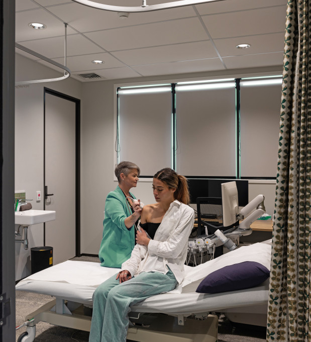
The skilled sonographer performing the examination will ask you questions about your scan, listen and make you comfortable. The procedure will be explained in detail and the sonographer will answer any questions you have before starting the examination.
Normally you will lie down on a bed and the area to be examined is exposed while the rest of the body is covered. Clear gel is applied to the area of your body which is being imaged. The sonographer will then place the “transducer” (a smooth hand held device) onto this area using gentle pressure. The transducer is moved across the area with a sliding and rotating action to allow the image to project onto the screen.
The sonographer takes still pictures from the images on the screen.
During the examination you may be asked to perform some simple actions to improve the quality of the imaging. For example, you may be asked to:
- “Take a breath” to assist during an abdominal ultrasound to allow the area beneath the rib cage to be clearly viewed.
- During an obstetric examination you may be asked to move to encourage the fetus or unborn baby to roll into a better position for imaging.
If any of these actions cause you concern or discomfort, you should let the sonographer know immediately.
Post Examination
You will be asked to change back into your clothes. Your scan will be interpreted by the Radiologist and a report will be sent directly to your doctor who will discuss the results with you.


