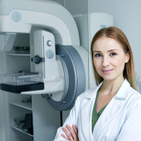
Breast MRI provides detailed images of internal and soft tissue structures of the breast, which enables a range of diagnostic procedures including assessment of implants, high risk screening and more accurate staging and treatment planning.
Non-invasive vascular studies using Doppler Ultrasound, CT scanning and MRI
Breast imaging includes techniques like mammography, ultrasound, and MRI to detect abnormalities, screen for cancer, and evaluate breast health.
A bone density scan measures bone mineral density using low-energy X-rays, helping to diagnose osteoporosis and assess fracture risk.
CT, or computed tomography, uses X-rays and computer processing to create cross-sectional images of the body for accurate diagnosis.
Interventional procedures utilize imaging guidance (like ultrasound, CT, or fluoroscopy) to perform diagnostic or therapeutic interventions.
MRI, or magnetic resonance imaging, utilizes powerful magnets and radio waves to produce detailed images of the body's internal structures.
Our state-of-the-art Molecular Imaging and Therapy Centre in Hastings
We are pleased to join you on your journey providing quality scanning to support the best outcome for you and your baby.
Ultrasound uses high-frequency sound waves to create real-time images of internal organs, tissues, and blood flow for diagnostic purposes.
X-ray imaging uses low-level radiation to produce images of the body's internal structures, aiding in diagnosis of fractures and diseases.

Part of Canopy Healthcare Group, Canopy Imaging (formerly TRG Imaging) was established in 2004 and is a leading provider of diagnostic imaging services.

Our focus is on ensuring expert imaging and diagnosis, with the leading sub-specialty skills of our team of Radiologists and over 300 staff.

We made it our mission to see our equipment through the eyes of our patients.

As part of our ongoing commitment to providing excellent customer care, we require more service orientated people to join our team.

Based at Auckland Breast Centre, we offer a 12-month intensive breast imaging fellowship.

Latest news and information from Canopy Imaging and the Canopy Healthcare Group
Breast MRI provides detailed images of internal and soft tissue structures of the breast, which enables a range of diagnostic procedures including assessment of implants, high risk screening and more accurate staging and treatment planning.
Before your Examination
Please phone us for an appointment. We will discuss:
Please let us know as soon as possible if there is any reason why you cannot keep your appointment.
Upon arrival:
You will have time to read about the examination, fill out a questionnaire as well as consent forms. In addition to a drink, insertion of an intravenous line may be necessary before your scan. A trained, experienced staff member will discuss the procedure with you. This is a good opportunity to ask any question you may have.
You will be asked to change into a gown and remove all metallic objects such as jewellery, dentures, hearing aids, etc.
Your Examination
If needed, you will have an intravenous line inserted. Our staff will position you on a bed that will gently move through the scanner, stopping to scan relevant areas. You will be asked to remain still throughout each of the 4-5 separate scans for up to twenty minutes. If you have difficulty lying flat, or are uncomfortable in confined spaces, the MRI staff will assist and make you more comfortable.
The Medical Radiation Technologist (MRT) or Radiographer will talk to you via a two-way intercom during the scan and let you know what is happening throughout. You can talk to the MRT at any time, even during the scan if you need to. If you have any questions, please don’t hesitate to ask. The MRI machine makes a loud tapping noise while scanning (from the MRI coil movement) however you will be able to listen to music during the scan.
Post Examination
You will be asked to change back into your clothes and your items will be returned to you. You will be monitored for 10 minutes. You may eat or drink after your scan. There are no known side effects from the magnetic field.
Your scan will be interpreted by the Radiologist and a report sent directly to your doctor who will discuss the results with you.